

Amniocentesis
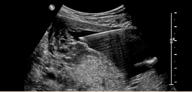
Amniocentesis is usually carried out in the 16th - 18th week of pregnancy. A very fine needle is inserted transabdominally and approx. 10 ml of fluid are removed from the amniotic sac. Local anesthesia is not required. The procedure usually lasts about 1 minute and is performed under constant ultrasound control.
Cells are cultured from the amniotic fluid for chromosomal analysis. The results are available about 10-14 days later. Upon request, the laboratory can carry out an additional rapid screening test (FISH, quick PCR), which gives a result after one or two days on the most common chromosomal abnormalities. In addition, the level of alpha-fetoprotein (AFP) in the amniotic fluid is determined. Elevated levels of AFP may indicate the development of spina bifida or an abdominal fissure. After genetic consultation, in special cases more tests can be performed for specific inherited diseases, such as metabolic disturbances. In rare cases, findings may be unclear, thus necessitating further examinations.
The risk of miscarriage from amniocentesis depends on the examiner and is usually given at around 0.25% - 1%. We recommend that patients go for a check-up examination with their regular gynecologist 1-2 days after the procedure, and that they avoid strenuous physical activity for a few days.
Chorionic villus sampling - Placental tissue biopsy
Chorionic villus sampling (biopsy of the placental tissue) is usually performed in the 12th - 14th week of pregnancy. During this procedure, cells from the placenta are removed transabdominally with a fine needle; ultrasound is used the whole time to visualize the procedure. As with amniocentesis, the procedure usually lasts about 1 minute.
The results of chromosomal analysis are available after 1-2 days (short-term culture) or after 8-10 days (long-term culture). In special cases, additional tests may be performed for inherited diseases, such as metabolic disturbances. In rare cases, findings may be unclear, thus necessitating further tests to be carried out.
The risk of miscarriage after chorionic villus sampling is approximately the same as for amniocentesis. We recommend that patients go for a check-up examination with their regular gynecologist 1-2 days after the procedure, and that they avoid strenuous physical activity for a few days.
Cordocentesis
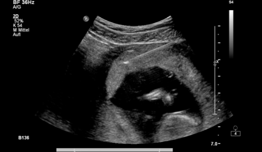
During cordocentesis, a small amount of fetal blood is taken from the umbilical vein. The procedure can be performed any time after the 18th week of pregnancy but, compared with amniocentesis and chorionic villus sampling, the risks involved are somewhat higher. The results of chromosomal analysis are available after only 2-3 days. Cordocentesis is still used to detect fetal infections and in the diagnosis of fetal blood disease.
Invasive fetal therapy
The unborn child can also be treated via injection into the umbilical vein with medication or blood products (intrauterine transfusion).
In rare cases, the puncture of fetal organs (e.g. bladder) or body cavities (abdominal cavity, chest) can aid diagnosis or therapy.